Personal or household handling, human administration, veterinary administration, and the use of laboratory preparation details as a dosing plan are strictly prohibited.
Oxytocin is a cyclic peptide made of nine amino acids. In laboratory studies, it is commonly used to examine the oxytocin receptor, usually shortened to OXTR.
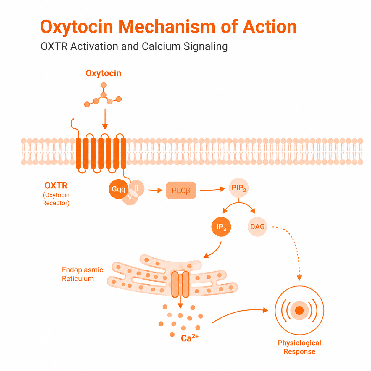
OXTR mainly signals through the Gq/11 pathway, which activates phospholipase C and raises calcium levels inside the cell. OXTR is the canonical receptor for oxytocin, but oxytocin is not universally selective for OXTR over vasopressin receptor subtypes.
The exact response can change with the cell type, receptor level, oxytocin concentration, exposure time, and test method.
Molecular Identity
Oxytocin has the sequence Cys-Tyr-Ile-Gln-Asn-Cys-Pro-Leu-Gly-NH2, also written as CYIQNCPLG-NH2. The C-terminal residue is glycinamide (Gly 9-NH2).
Its CAS number is 50-56-6, its molecular formula is C43H66N12O12S2, its average molecular weight is about 1,007.2 g/mol, and its monoisotopic mass is 1,006.4365 Da.
A disulfide bond joins Cys1 and Cys6.[1]
This formula, average molecular weight, and monoisotopic mass describe the neutral oxytocin molecule. Commercial research materials may be supplied as free peptide, acetate salts, or hydrates.
Molar stock calculations based on weighed material should account for the batch-specific peptide content, counterion, and water content.
The average molecular weight of 1,007.2 g/mol should not be used as the exact high-resolution mass spectrometry (HRMS) target; the expected HRMS result depends on whether the report gives the neutral monoisotopic mass (1,006.4365 Da), a protonated ion ([M+H]+ approximately 1,007.444 m/z), a multiply charged ion (e.g., [M+2H]2+ approximately 504.226 m/z), an adduct, or a salt-associated species.
The disulfide bond creates the ring-shaped part of the peptide. The last three residues form a short tail.
A cryo-electron microscopy structure of oxytocin bound to active human OXTR (PDB 7QVM, Waltenspühl et al., Nat Commun 2022) shows that both the ring and the tail make contacts with the receptor. Disruption of the disulfide bond or mutation of key contact residues changes receptor binding and signaling.[2]
How OXTR Starts the Signal
Human OXTR, represented by UniProt entry P30559, is a 389-amino-acid class A G-protein-coupled receptor.[3]
In many OXTR-expressing cell systems, oxytocin binding activates Gq/11. Gq/11 then activates phospholipase C-β, which splits phosphatidylinositol 4,5-bisphosphate into IP3 and diacylglycerol.
IP3 releases Ca2+ from intracellular stores, while diacylglycerol helps activate protein kinase C.[4][5]
OXTR can also connect to Gi/o, Gs, and other signaling proteins in some test systems. These extra pathways are not seen in the same way in every model.
Researchers should measure the pathway in the chosen cells instead of assuming that OXTR always produces one fixed response.[4]
Calcium Signaling
Oxytocin can produce a short calcium rise, a longer response, or repeated calcium spikes in cultured cell models. The pattern depends on the cell preparation, receptor level, concentration range, extracellular calcium, and recording method.[6]
In one study of cultured human myometrial cells, oxytocin at 10–300 nM typically induced calcium oscillations at approximately 0.6–0.8 cycles per minute. Within that concentration range, neither oscillation frequency nor amplitude changed
significantly.
This finding should not be generalized to other cell models or concentration ranges.[7]
Desensitization and Receptor Recycling
OXTR signaling can become weaker during continued receptor activation. In cell studies, activated OXTR can recruit G-protein-coupled receptor kinases and β-arrestins.
These proteins reduce further G-protein signaling and help move the receptor away from the cell surface. GRK2 and β-arrestin recruitment can begin within seconds—GRK2 interaction has been detected from approximately 4 seconds and β-arrestin
from approximately 10 seconds after receptor activation in a receptor-expression model.[8]
An internalized receptor is not always destroyed. In one cultured-cell model, OXTR returned to the cell surface through a Rab4/Rab5 recycling route.
Other models may show different timing or a different balance between recycling and breakdown. Receptor trafficking should therefore be measured in the actual test system.[9]
Biased Signaling and Other Pathways
Some oxytocin analogs activate one OXTR pathway more strongly than another. This is often called biased signaling.
For example, an analog may give a different balance of G-protein signaling, calcium release, β-arrestin recruitment, receptor internalization, or receptor recycling.[10]
The words “balanced” and “biased” should only be used when the study states the reference ligand, the pathways being compared, the cell system, and the calculation method.
A single calcium or ERK result is not enough to prove that a ligand is biased.
OXTR activation can also change ERK1/2 and other downstream signals in some cell models. These are pathway measurements, not automatic proof of changes in growth, differentiation, survival, contractility, or behavior.
A biological claim needs a direct test of that outcome in the same model.[4]
Receptor Selectivity
Oxytocin primarily targets OXTR, but it can also interact with vasopressin receptor subtypes in some assays. Reported affinity and potency values change with the receptor subtype, species, expression system, radioligand, incubation conditions, and test
endpoint.
For this reason, a fixed selectivity ratio such as “100:1” should not be used as a universal value.[11]
When an experiment uses concentrations that may activate more than one receptor subtype, receptor-negative controls, receptor knockdown, or suitable subtype-selective controls can help show which receptor produced the result.
Laboratory Handling and Stability
- Storage: Follow the batch label, certificate of analysis, and product stability document. When the batch instructions specify frozen storage, keep the sealed lyophilized material at about −20°C, protected from light
and moisture. Avoid repeated warming and cooling. - Reducing agents: Do not add DTT, β-mercaptoethanol, or another reducing agent unless disulfide reduction is the purpose of the experiment.
- Solubility: Oxytocin is listed as soluble in water, but the usable concentration can still change with pH, buffer salts, counterion, peptide content, and batch condition.[1]
- Laboratory preparation only: Choose the
solvent and concentration for the stated analytical or cell-assay method. References to water or buffered saline describe laboratory pipetting only and do
not describe a preparation for administration. - Prepared solutions: Refrigeration alone does not guarantee stability. Oxytocin can undergo deamidation, oxidation, hydrolysis, aggregation, and thiol-disulfide exchange. Stability can change with pH, temperature, light, oxygen,
metal ions, container type, concentration, and storage time.[12]
How to Read a Research COA
A certificate of analysis is a batch quality record. It does not show that the material is suitable for every experiment.
HPLC and mass spectrometry answer different questions:
- HPLC: shows the relative amount of peaks detected by that chromatographic method. The result depends on the column, mobile phase, gradient, detector, wavelength, integration rules, and sample preparation.
- Mass spectrometry: can show that the observed mass matches the expected peptide. The expected result depends on whether the report uses neutral monoisotopic mass (1,006.4365 Da), a protonated ion ([M+H]+ approximately
1,007.444 m/z), a multiply charged ion ([M+2H]2+ approximately 504.226 m/z), or a deconvoluted neutral mass. A matching intact mass supports identity, but it does not by itself prove correct disulfide pairing, purity, peptide content,
sterility, or biological activity. - Peptide content: is not the same as HPLC area purity. Water, counterions, residual solvents, and non-peptide material can reduce the amount of peptide in a weighed sample even when the main HPLC peak is high.
A useful research COA may include the batch number, appearance, identity result, HPLC method and purity result, observed mass, peptide content when measured, water, counterion, residual solvents, related peptide impurities, and storage conditions. Which
tests are needed depends on the synthesis process and the planned laboratory assay.
The comprehensive characterization described in published peptide reference-standard literature (e.g., multi-laboratory assigned values, mass balance with water and counterion) represents a high-level reference approach; a routine RUO COA may contain
fewer tests depending on product grade and intended use.[13]
ICH Q2(R2) is a pharmaceutical analytical-validation framework rather than a universal requirement for RUO COAs. Its fit-for-purpose principles—including selectivity, accuracy, precision, range, and robustness—are useful when evaluating analytical
reliability.[14]
For animal studies, primary-cell assays, or immune-sensitive endpoints, endotoxin and, where relevant, bioburden or sterility testing may also be required. A COA that meets its stated limits confirms only the listed batch tests.
It does not prove safety or effectiveness for human, veterinary, cosmetic, or other non-research use.
Basic Study Design
- Record the batch number, peptide form, stated purity, observed mass, storage history, solvent, stock concentration, final assay concentration, and exposure time.
- Use a concentration-response series and a time course instead of relying on one concentration and one reading.
- Include vehicle controls and, where suitable, receptor-negative, receptor-knockdown, or receptor-blocking controls.
- Separate pathway measurements from biological outcomes. Calcium, IP3, ERK, β-arrestin recruitment, and receptor internalization do not by themselves prove a change in a larger cell or animal outcome.
- Check for vasopressin-receptor cross-reactivity when the concentration or model makes it relevant.
Research Use Only Disclaimer
Research Use Only: All NEXALUME LABS products are intended and labeled solely for laboratory research use. They are not intended, manufactured, tested, or released for human or veterinary administration.
This article provides general scientific information regarding standard laboratory in vitro and in vivo research contexts only, and does not constitute a validated experimental protocol, medical guidance, clinical guidance, veterinary guidance, or treatment
advice.