Research Use Only Notice: This article is about Kisspeptin-10 as a laboratory research reagent. It only covers in vitro tests, ex vivo tissue work, and laboratory animal studies conducted under institutionally approved protocols.
It is not intended or approved for human consumption, administration, diagnosis, treatment, cosmetic use, personal use, or veterinary use. Laboratory staff should follow the product label, batch Certificate of Analysis (COA), Safety Data Sheet (SDS), and their approved protocol.
Species note: The molecular data in this article refer to human Kisspeptin-10. Rodent KP-10 sequences differ—for example, rat and mouse KP-10 is YNWNSFGLRY-NH2 (position 10 is Tyr, not Phe), with a different molecular formula, molecular weight, and CAS number.
Researchers should confirm the peptide identity against the actual species sequence and batch COA before cross-species experiments.
Results from one test system should not be treated as proof of the same effect in another species, tissue, or experiment. This article therefore keeps cell, tissue, and animal findings clearly separated.
Molecular Profile
The amino acid sequence of human Kisspeptin-10 is Tyr-Asn-Trp-Asn-Ser-Phe-Gly-Leu-Arg-Phe-NH2, also written as YNWNSFGLRF-NH2. PubChem lists its molecular formula as C63H83N17O14, its average molecular weight as about 1,302.4 g/mol (non-salt human peptide moiety), its monoisotopic mass as 1,301.6305 Da, and its CAS Registry Number as 374675-21-5.[3]
| Property | Reference Information |
|---|---|
| Sequence | YNWNSFGLRF-NH2 (human) |
| Length | 10 amino acids |
| Molecular formula (non-salt human peptide) | C63H83N17O14 |
| Average molecular weight | About 1,302.4 g/mol (non-salt human peptide moiety) |
| Monoisotopic mass (non-salt human peptide) | 1,301.6305 Da[3] |
| CAS number | 374675-21-5 |
| Common laboratory form | Lyophilized synthetic peptide; the supplied salt form (e.g., TFA or acetate) and peptide content may differ by batch. The molecular weight used for molar concentration calculations should reflect the actual salt form and batch-specific peptide content as stated on the COA. |
KP-10 is the C-terminal 10-amino-acid part of kisspeptin-54 and is the smallest kisspeptin fragment commonly described as retaining strong KISS1R agonist activity. Structure-activity studies found that substitutions at key positions, particularly residues 6 and 10, can greatly reduce functional potency in tested receptor systems.[5]
These results apply to the exact analogues and assays that were tested and should not be turned into a claim that every terminal change always removes all activity.
KP-10 may show receptor activity similar to longer kisspeptins in some cell assays, but that does not mean it has the same stability or duration in an animal model. A study of modified KP-10 analogues with enhanced resistance to enzymatic breakdown showed improved in-vivo activity compared with native KP-10.[4]
The average molecular weight of 1,302.4 g/mol refers to the non-salt human peptide moiety and should not be used directly for molar calculations if the supplied batch contains TFA, acetate, water, or other counterions. For example, a 1:1 TFA salt form has an approximate nominal molecular weight of about 1,416.5 g/mol.
The batch-specific molecular weight, counterion result, and peptide-content value from the COA should be used for solution preparation.
How Kisspeptin-10 Activates KISS1R
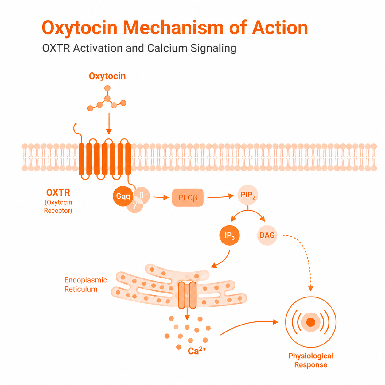
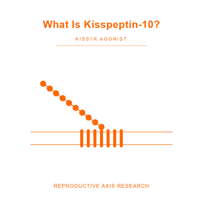
KISS1R is a G-protein-coupled receptor. In receptor-expressing cell systems, activation of KISS1R can turn on the Gq/11-PLC pathway.
This produces IP3 and DAG signals, raises intracellular calcium, and can activate PKC and other downstream pathways.[1][2]
The exact response depends on the cell type, receptor level, peptide concentration, test method, and measurement time. For this reason, Kd, Ki, IC50, and EC50 are not interchangeable values.
A binding result should not be reported as a functional potency result, and a result from one assay should not be presented as a fixed property of every KP-10 experiment.
Babwah et al. (2012) used single-cell analyses of KISS1R-expressing cells and found both a rapid intracellular calcium rise and a longer sustained calcium response that depended on calcium entering from outside the cell.[6]
Zhang et al. (2008) showed in ex vivo mouse brain-slice studies that kisspeptin increased the activity of GnRH neurons mainly through TRPC-like cation channels, with a smaller contribution from reduced inward-rectifier potassium current.[7]
This means the response should not be explained as calcium influx alone.
What Research Models Show
The kisspeptin pathway is important in reproductive neuroendocrine research, but it should not be called the only “master switch” or the whole GnRH pulse generator.
Wakabayashi et al. (2013) used bilateral electrodes targeting the arcuate KNDy region in goats and recorded synchronized multiunit activity volleys; anatomical studies supported bilateral connections among neurokinin-B-containing neurons. The study supports a network model involving several signals (kisspeptin, neurokinin B, and dynorphin) rather than a single peptide acting alone.[8]
Mouse studies also show that sex-steroid changes can affect Kiss1 gene expression differently in different brain regions. Smith et al. (2005) found that estrogen-related changes in the arcuate nucleus differed from those in the anteroventral periventricular region.[9]
These findings are specific to the mouse model and do not show that kisspeptin neurons are the only place where steroid feedback occurs.
Common Laboratory Uses
- Receptor assays: KP-10 can be used as an agonist control in cells engineered to express KISS1R. Common readouts include calcium signals, phosphoinositide signaling, receptor movement, and concentration-response curves.
- Cell-signaling studies: Researchers may compare KP-10 responses with vehicle controls, receptor-negative cells, pathway blockers, or genetically changed cells.
- Ex vivo tissue studies: Approved brain-slice experiments may measure membrane voltage, firing rate, ion-channel activity, or calcium changes. Freshly isolated tissue or acute slices are ex vivo; cultured primary cells are in vitro.
- Laboratory animal models: Approved studies may use KP-10 to examine pathway responses. Hormone measurements are downstream readouts of pathway activity and do not by themselves demonstrate direct KISS1R engagement or a specific cellular mechanism. They must not be described as diagnostic tests.
Each study should clearly report the species or cell line, tissue source, receptor construct, solvent, concentration, exposure time, test method, controls, and statistical method. Without these details, results from different experiments cannot be compared reliably.
How to Read a Kisspeptin-10 COA
A COA applies to one tested batch. It does not prove that every batch with the same product name has the same purity, salt form, peptide content, water level, or residual solvent profile.
| COA Test | What It Shows | What It Does Not Show by Itself |
|---|---|---|
| HPLC or UPLC purity | The share of the recorded chromatographic signal assigned to the main peak under the stated method. The result is detector- and method-dependent; different impurities may have different UV response factors, so area percentage does not necessarily equal mass percentage. | Full sequence identity, exact peptide content, sterility, or biological activity |
| Mass spectrometry | Whether the measured ion or ions match the expected molecular mass | Complete purity or the amount of peptide in the weighed powder |
| Water test | Water content under the stated test method | Peptide identity or activity |
| Residual solvent test | The amount of the solvents included in the test panel | Absence of solvents that were not tested |
| Counter-ion test | The amount of a named counter-ion, such as TFA or acetate, when tested | Net peptide content unless the COA also gives a full content calculation |
HPLC and mass spectrometry should be read together: HPLC mainly describes the chromatographic peak pattern, while mass spectrometry supports molecular identity. A useful COA should also list the batch number, test method or method identifier, result, acceptance limit, test date, and approval status.
There is no single purity requirement for every KP-10 study. A laboratory may set a purchase specification such as ≥95% or ≥98% by HPLC, depending on the work, but the actual batch result must come from the COA.
ICH Q2(R2) explains that an analytical method should be suitable for its intended purpose and should have documented performance, including suitable selectivity, accuracy, and precision. ICH Q2(R2) is a pharmaceutical analytical-validation guideline; its principles may be used as a technical reference for research-use-only peptide methods, but it does not automatically establish mandatory release requirements for every RUO product.[10]
Residual solvent testing should focus on solvents that may remain from manufacturing or purification. ICH Q3C(R9) is a useful technical reference for solvent classes and analytical testing, but it is written for pharmaceutical quality systems and does not automatically create a universal release limit for every research-use-only peptide.[11]
COA Limit: A passing COA only shows that the tested batch met the listed laboratory specifications. It does not prove safety, clinical effect, sterility, endotoxin status, or suitability for human, cosmetic, or veterinary use.
Storage and Laboratory Solution Preparation
Laboratory Preparation Only: The information below is only for trained laboratory staff preparing research solutions. It is not a human or veterinary reconstitution, dosing, administration, or injection guide.
- Lyophilized material: A common laboratory starting condition is −20°C, dry, tightly closed, and protected from light. The batch label and COA always take priority.
- Solvent: Use the solvent listed in the batch documentation or validated in the laboratory protocol. Do not assume that every salt form or batch has the same water solubility.
- Concentration: Use the verified net peptide content, the correct molecular mass for the salt form and species, and not only the total powder weight when preparing a molar stock.
- Aliquots: Where the study protocol allows it, divide the solution into small, labeled aliquots to reduce repeated freeze-thaw cycles.
- Hold time: Do not assign a general refrigerated shelf life unless the exact solvent, concentration, container, temperature, and test method have been studied.
KP-10 stability changes with the sample matrix. Liu et al. (2013) reported rapid loss of intact KP-10 and formation of a principal degradation product in rat plasma; the exact structural assignment should be taken from the original analytical data.[12]
This result is useful for rat-plasma experiments, but it does not set the shelf life of a laboratory stock solution in another solvent.
Research References
- Ohtaki T, Shintani Y, Honda S, et al. Metastasis suppressor gene KiSS-1 encodes peptide ligand of a G-protein-coupled receptor. Nature. 2001;411(6837):613-617. doi:10.1038/35079135. PubMed.
- Kotani M, Detheux M, Vandenbogaerde A, et al. The metastasis suppressor gene KiSS-1 encodes kisspeptins, the natural ligands of the orphan G protein-coupled receptor GPR54. Journal of Biological Chemistry. 2001;276(37):34631-34636. doi:10.1074/jbc.M104847200. PubMed.
- National Center for Biotechnology Information. PubChem Compound Summary for CID 25240297, Kisspeptin-10 (human). PubChem.
- Curtis AE, Cooke JH, Baxter JE, et al. A kisspeptin-10 analog with greater in vivo bioactivity than kisspeptin-10. American Journal of Physiology-Endocrinology and Metabolism. 2010;298(2):E296-E303. doi:10.1152/ajpendo.00426.2009. PubMed.
- Gutiérrez-Pascual E, Leprince J, Martínez-Fuentes AJ, et al. In vivo and in vitro structure-activity relationships and structural conformation of Kisspeptin-10-related peptides. Molecular Pharmacology. 2009;76(1):58-67. doi:10.1124/mol.108.053751. PubMed.
- Babwah AV, Pampillo M, Min L, Kaiser UB, Bhattacharya M. Single-cell analyses reveal that KISS1R-expressing cells undergo sustained kisspeptin-induced signaling that is dependent upon an influx of extracellular Ca2+. Endocrinology. 2012;153(12):5875-5887. doi:10.1210/en.2012-1615. PubMed.
- Zhang C, Roepke TA, Kelly MJ, Rønnekleiv OK. Kisspeptin depolarizes gonadotropin-releasing hormone neurons through activation of TRPC-like cationic channels. Journal of Neuroscience. 2008;28(17):4423-4434. doi:10.1523/JNEUROSCI.5352-07.2008. PubMed.
- Wakabayashi Y, Yamamura T, Sakamoto K, Mori Y, Okamura H. Electrophysiological and morphological evidence for synchronized GnRH pulse generator activity among kisspeptin/neurokinin B/dynorphin A (KNDy) neurons in goats. Journal of Reproduction and Development. 2013;59(1):40-48. doi:10.1262/jrd.2012-136. PubMed.
- Smith JT, Cunningham MJ, Rissman EF, Clifton DK, Steiner RA. Regulation of Kiss1 gene expression in the brain of the female mouse. Endocrinology. 2005;146(9):3686-3692. doi:10.1210/en.2005-0488. PubMed.
- International Council for Harmonisation. ICH Q2(R2): Validation of Analytical Procedures. 2023. ICH.
- International Council for Harmonisation. ICH Q3C(R9): Impurities: Guideline for Residual Solvents. 2024. ICH.
- Liu Z, Ren C, Jones W, et al. LC-MS/MS quantification of a neuropeptide fragment kisspeptin-10 and characterization of its decomposition product and pharmacokinetics in rats. Journal of Chromatography B. 2013;926:1-8. doi:10.1016/j.jchromb.2013.02.027. PubMed.
Research Use Only Disclaimer
Research Use Only: All NEXALUME LABS products are intended and labeled solely for laboratory research use. They are not intended, manufactured, tested, or released for human or veterinary administration.
This article provides general informational context concerning in vitro, ex vivo, and laboratory-animal research; it is not a validated protocol or standard operating procedure, and does not constitute medical, clinical, veterinary guidance, or treatment advice.